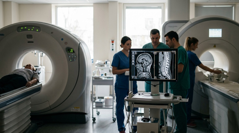
CT and MRI are the two most powerful diagnostic tools available, yet they are frequently confused. While both produce cross-sectional images of your internal organs, they operate on entirely different physical principles. Choosing the right method is critical for an accurate diagnosis: what shows up clearly on a CT scan might be invisible on an MRI, and vice versa.
So, what exactly is the difference, and which one should you choose?
Imagine a doctor needs to look inside your body.
- One way is to use X-rays from multiple angles to create “slices” of your anatomy—that’s a CT scan.
- The other way is to use a powerful magnetic field and radio waves to make the hydrogen atoms in your tissues “respond,” building an image from those signals—that’s an MRI.
In simple terms, a CT is an advanced, 3D version of an X-ray. An MRI is a completely different technology that uses no ionizing radiation. Because of this, they produce different results: CT is superior for dense structures like bones, acute bleeding, and the lungs, while MRI excels at visualizing soft tissues like the brain, ligaments, and spinal discs.
The Fundamental Difference: X-rays vs. Magnetic Fields
The core distinction lies in the physics behind the machines.
- Computed Tomography (CT) uses X-ray radiation. An X-ray tube rotates around the patient, while sensors capture the beams as they pass through the body. The denser the tissue, the fewer rays pass through, which creates the image.
- Magnetic Resonance Imaging (MRI) works differently: it generates a massive magnetic field that aligns the hydrogen protons in your body. Radiofrequency pulses then “excite” these protons. As they return to their original state, they emit faint signals that the computer captures and converts into a picture. This makes MRI much safer for repeated studies since there is no exposure to ionizing radiation.
What CT Sees Best vs. What MRI Sees Best
CT scans are indispensable for evaluating bone tissue: fractures, cracks, osteoporosis, and bone infections are perfectly visible. CT is also the gold standard for imaging the lungs (pneumonia, tumors, tuberculosis), detecting internal bleeding (fresh blood appears bright on a CT), identifying calcifications, and finding kidney or gallbladder stones. It is the go-to method for trauma and strokes (to rule out a brain bleed) because every second counts in an emergency.
MRI, on the other hand, is the ultimate tool for soft tissues. It provides flawless visualization of the brain and spinal cord, intervertebral discs, ligaments, tendons, cartilage, muscles, and pelvic organs. MRI is essential when a doctor suspects multiple sclerosis (MS), brain tumors, herniated discs, or tears in the meniscus or ACL. Additionally, contrast-enhanced MRI is superior for detecting inflammatory and demyelinating diseases.
To help visualize these differences, let’s look at this comparison table:
| Feature | Computed Tomography (CT) | Magnetic Resonance Imaging (MRI) |
| Principle | X-ray radiation | Magnetic field + Radio waves |
| Best For | Bones, lungs, bleeding, stones | Brain, ligaments, discs, cartilage |
| Risk Factor | Ionizing radiation exposure | Safe, no radiation; repeatable |
| Duration | 5–15 minutes | 20–60 minutes or longer |
| Contraindications | Pregnancy, childhood (relative) | Metal implants, pacemakers, claustrophobia |
When a CT Scan is Ordered: Specific Scenarios
CT is most often used in emergency situations.
It is the first line of defense for severe trauma—when doctors need to quickly assess fractures of the skull, spine, or pelvis. A CT is also performed if a hemorrhagic stroke is suspected, as it is the best method for finding blood in the brain.
Furthermore, CT is used to diagnose pneumonia, tuberculosis, and lung cancer (often as a screening for heavy smokers). For abdominal pain, a CT scan helps locate kidney stones, gallstones, abscesses, or abdominal tumors. When it comes to checking the sinuses for chronic sinusitis, CT remains the “gold standard.”
When an MRI is Ordered: The Preferred Choice
MRI is usually a scheduled procedure where there is no immediate emergency, but a detailed view of soft tissue is required.
Typical cases include:
-
Unexplained headaches, dizziness, or suspected tumors/Multiple Sclerosis (Brain MRI).
-
Back pain radiating down the leg—a spinal MRI will show herniated discs or spinal stenosis.
-
Knee or shoulder injuries—a joint MRI can detect torn ligaments, tendons, or menisci.
-
Hip joint issues (such as avascular necrosis).
Additionally, contrast-enhanced MRI is vital for diagnosing inflammatory bowel diseases like Crohn’s and for evaluating oncological progression.
Contraindications and Limitations: What You Need to Know
CT has virtually no absolute contraindications, except for pregnancy (due to radiation risk to the fetus). In children, CT is only performed when strictly necessary.
MRI, however, has several absolute contraindications: the presence of any ferromagnetic metal in the body (implants, shrapnel, vascular clips), pacemakers, insulin pumps, cochlear implants, or non-removable braces. MRI can also be challenging for those with claustrophobia (fear of enclosed spaces), potentially requiring sedation or the use of an “open” MRI machine.
Unlike the quick CT, an MRI takes much longer and requires the patient to remain perfectly still; if a patient is in severe pain or coughing uncontrollably, a CT may be substituted.
Additional Factors: Speed and Contrast
The speed of the study is a major factor.
A CT is finished in just a few minutes, which is critical for trauma and intensive care. An MRI can take anywhere from 30 minutes to an hour.
- When using contrast dyes, CT utilizes iodine-based agents (which can cause allergies or strain the kidneys).
- MRI contrast is gadolinium-based; it is less likely to cause an allergic reaction and is less toxic to the kidneys, though its long-term accumulation in the brain after frequent use is a topic of ongoing debate.
The final choice of method is always made by your physician based on the clinical picture, the suspected diagnosis, and your overall health. Never attempt to self-diagnose: what feels like “just a pinched nerve” might require a spinal MRI, while a head injury always requires an immediate CT.
Welcome to Poznayu.com!
My name is Alex, and I founded this project together with a team of like-minded professionals. At Poznayu.com, we create in-depth reviews, explore fascinating facts, and share well-researched, reliable knowledge that helps you navigate complex topics with confidence.
Our mission is simple: to explain complicated ideas in clear, accessible language. We believe that high-quality information should be available to everyone. Every article we publish is designed to provide practical value, actionable insights, and trustworthy analysis you can rely on.
Join our growing community of curious readers. Your feedback matters — share your thoughts in the comments, ask questions, and suggest topics you’d like us to cover next.