Osteoarthritis (OA) is a chronic degenerative joint disease characterized by the gradual breakdown of cartilage.
Contrary to popular belief, it is not simply mechanical “wear and tear” caused by aging. It is a complex pathological process that affects all components of the joint.
- Cartilage loses elasticity, becomes thinner, and develops cracks, after which the adjacent bone, synovial lining, ligaments, and surrounding muscles become involved in the pathological process.
- The disease is accompanied by altered metabolism within the joint capsule, leading to chronic inflammation and remodeling of bone structures with the formation of osteophytes — bony spurs.
This condition is the most common cause of joint pain worldwide and the leading threat to mobility in people over forty.
Osteoarthritis most often affects large weight-bearing joints that take on the greatest loads: the knees (gonarthrosis) and hips (coxarthrosis).
Shoulder osteoarthritis (omarthrosis) is less common than knee or hip osteoarthritis because the shoulder does not bear the body’s axial weight:
- It most often develops secondarily due to old injuries, recurrent dislocations, rotator cuff damage, or regular heavy overhead pressing.
- The disease causes deep pain when raising the arm overhead, crepitus, and severe restriction of rotation, making timely MRI evaluation necessary.

This is especially interesting for athletes: how can you distinguish the symptoms of shoulder osteoarthritis from, for example, bursitis or rotator cuff pain?
In very simple terms, osteoarthritis causes deep, chronic pain, crepitus, and the same restriction of both active and passive movement in all planes (clearly visible on X-rays as joint-space narrowing and osteophytes).
- Bursitis is characterized by sharp, localized pain with swelling when the arm is abducted through 60–120° and at night, while passive joint motion remains completely preserved (diagnosed by ultrasound).
- Rotator cuff damage causes loss of strength: the patient cannot actively raise or hold the arm because of a tendon tear, although a physician can easily raise it passively (confirmed by MRI).
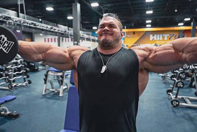
Interestingly, tremendous strength does not rule out degenerative changes in a joint. In my own practice, for example, I have seen athletes who, after warming up, could perform 12 lateral raises with 88-pound dumbbells. Yet even that level of strength did not necessarily protect them from degenerative joint changes.
For example,
- If this is Stage 1 or early Stage 2 (shoulder osteoarthritis): the cartilage has already begun to lose elasticity and thin out, and the first osteophytes appear, but there is still no anatomical (“bony”) block. The joint space is not critically narrowed, so movement biomechanics remain preserved.
- Acromioclavicular joint (AC joint) osteoarthritis: this is a classic condition among strength athletes (“weightlifter’s shoulder”). It is not the glenohumeral joint itself that is damaged, but the small acromioclavicular joint above it. During lateral raises for the middle deltoid, strength may not decrease at all; the disease shows up only as localized pinpoint pain when the clavicle is palpated and an aching discomfort after training.
However, the disease can also affect the small joints of the hands and feet and the facet joints of the spine.
The insidious nature of the disease lies in its irreversibility — modern medicine can slow or stop cartilage degradation, but conservative methods cannot restore tissue that has already been destroyed.
Osteoarthritis Symptoms and Clinical Presentation
In its early stages, osteoarthritis develops almost without symptoms, because cartilage itself contains no nerve endings or pain receptors.
The first signs of disease appear only when the pathological process involves the synovial lining or bone tissue. Patients usually notice mild discomfort after prolonged physical activity, extended walking, or carrying heavy loads.
A characteristic early marker is “start-up pain” — brief joint stiffness after waking up or sitting for a long time that subsides after a few minutes of movement as synovial fluid redistributes across the cartilage surface.
As the disease progresses, symptoms become more pronounced and persistent.
The pain takes on a mechanical pattern, becoming worse when weight is placed on the affected limb and easing at rest. As the cartilage cushioning layer thins, the joint surfaces begin rubbing against each other without adequate shock absorption, producing characteristic coarse crepitus. At the same time, the surrounding muscles undergo reflex spasm, which the body uses as a protective mechanism to immobilize the damaged area, inevitably restricting range of motion.
In later stages, the clinical picture is compounded by visible anatomical changes.
The joint becomes deformed because of marginal osteophyte growth and thickening of the joint capsule. Reactive synovitis may occur periodically — acute inflammation of the inner lining with excessive accumulation of pathological fluid, causing visible swelling, localized tissue warmth, and acute pain even without weight-bearing or exertion.
The main clinical manifestations that indicate the development of the condition include:
- Mechanical pain directly related to physical activity and disappearing during prolonged rest.
- Morning joint stiffness, typically lasting from 15 to 30 minutes.
- Reduced range of motion, with an inability to fully bend or straighten the affected limb.
- Specific dry, coarse crepitus during movement, always accompanied by pain.
- Visible changes in the joint contour, with dense swelling or hard bony deformities.
Modern Diagnostic Methods for Osteoarthritis
Osteoarthritis is diagnosed and treated by orthopedic and trauma specialists, while a rheumatologist becomes involved when systemic autoimmune processes are suspected.
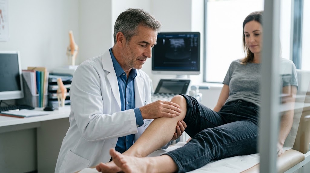
The initial visit always begins with a medical history and a thorough physical examination. The physician palpates the joint, assesses symmetry of the affected area, checks active and passive range of motion, and performs functional stress tests. Manual examination allows the specialist to identify the precise location of pain, the presence of an effusion (excess fluid) within the joint capsule, and the degree of muscle atrophy that often accompanies longstanding degenerative processes.
Conventional X-ray remains the gold standard for imaging diagnosis.
Images are taken in two views, and knee X-rays require weight-bearing imaging while standing. Cartilage itself is not visible on an X-ray, but the physician evaluates joint-space height — the distance between the contacting bones. X-rays show marginal bone growths (osteophytes), subchondral sclerosis (pathological thickening of the bone beneath the cartilage), and possible cystic changes very clearly. The radiographic stage of disease is then assigned according to the international Kellgren-Lawrence classification, which determines the subsequent treatment protocol.
Magnetic resonance imaging (MRI) is used when detailed evaluation of soft tissues that cannot be visualized with X-rays is required.
MRI provides detailed, slice-by-slice visualization of hyaline cartilage, the menisci, ligaments, the joint capsule, and the synovial lining. MRI is indispensable in the early stages (pre-osteoarthritis), when X-rays still do not show joint-space narrowing but hidden dehydration processes have already begun within the cartilage, or when bone marrow edema has developed within the cancellous bone.
Ultrasound is used for ongoing assessment of soft-tissue structures.
This method is inferior to MRI for visualizing deep intra-articular structures, but it is well suited for detecting synovitis, assessing cartilage thickness in accessible areas, and providing precise guidance for intra-articular injections. Laboratory testing plays a supporting role in isolated osteoarthritis.
A complete blood count, blood chemistry, C-reactive protein testing, rheumatoid factor, and uric acid testing are ordered solely to rule out rheumatoid arthritis, gout, or infectious disease requiring a fundamentally different treatment approach.
Injection Therapy: Medications and Treatment Protocols
Injection therapy is selected individually based on the current stage of disease and the presence of active inflammation.
Choosing the right medication — from emergency anti-inflammatory injections to restorative “liquid prostheses” — can rapidly control pain and protect cartilage, especially when procedures are performed under ultrasound guidance.
Chondroprotectors and Local Anesthetics
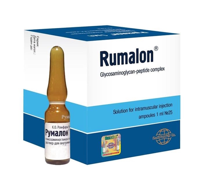
Preparations based on extracts of animal cartilage and bone marrow, such as Rumalon, contain active glycosaminoglycan-peptide complexes.
They are used to stimulate cartilage regeneration, improve the quality of the body’s own synovial fluid, and slow destructive enzymes. Because deep intramuscular injection of such dense solutions often causes significant pain, their use in clinical practice is combined with local anesthetics such as lidocaine. Lidocaine rapidly blocks sodium channels in nerve endings, preventing transmission of pain impulses and making the procedure more comfortable.
This therapeutic combination is used primarily during the first and second stages of osteoarthritis, strictly outside the acute inflammatory phase. The injection course has a prolonged cumulative effect, so immediate restoration of joint structure should not be expected. In this protocol, the anesthetic serves the practical purpose of reducing injection pain, while the chondroprotector provides long-term nutritional support for the depleted cartilage matrix.
Glucocorticoids With Anesthetics (Therapeutic Injections)
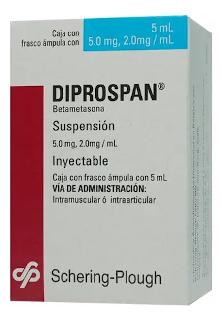
When reactive synovitis develops and the joint becomes swollen, hot to the touch, and extremely painful, using structural preparations no longer makes sense.
In these situations, orthopedists use intra-articular injections of potent hormonal anti-inflammatory drugs, such as Diprospan or Kenalog, combined with lidocaine, ropivacaine, or bupivacaine. The anesthetic immediately “switches off” the pain, allowing the patient to put weight on the leg right after the procedure, while the synthetic glucocorticoid suppresses the immune response for several days, reduces tissue swelling, and interrupts the inflammatory cascade.
A corticosteroid injection is an emergency measure intended solely to relieve acute symptoms, not to treat osteoarthritis systemically. These hormones have a pronounced catabolic effect: when injected too often, they disrupt protein metabolism in the joint and dramatically accelerate the degradation of already damaged cartilage. For this reason, protocols strictly limit the number of such injections (no more than three injections into one joint per year), using them only to move the disease from an acute phase into stable remission.
Hyaluronic Acid Preparations (Liquid Prostheses)
Hyaluronic acid is a critically important component of synovial fluid, responsible for its natural viscosity and lubricating properties.
In Stage 2 or Stage 3 osteoarthritis, the joint’s natural lubrication loses its physical and chemical properties, becomes excessively watery, and stops protecting cartilage from mechanical friction. Injecting high-density synthetic hyaluronic acid directly into the joint capsule can create a lasting effect of artificial biomechanical cushioning.
Liquid prostheses should be injected only into a “clean” and calm joint, meaning after all signs of acute synovitis have been eliminated; otherwise, inflammatory exudate will rapidly dissolve and destroy the expensive preparation. The thick gel coats the joint surfaces with a durable protective film, mechanically separates the contacting bones, and dramatically reduces pain while walking.
The effect of a course of injections lasts from six months to one and a half years, which can significantly delay the need for major surgery.
Plasma Therapy (PRP Therapy)
Platelet-rich plasma therapy (PRP) is a biotechnology-based method that uses the body’s own regenerative resources.
The patient provides venous blood, which is centrifuged to isolate plasma with an extremely high concentration of platelets. The resulting autologous preparation is then injected into the affected joint. When platelets come into contact with damaged tissue, they degranulate and release powerful growth factors that activate natural repair cascades, stimulate the formation of new microcapillaries, and gently suppress low-grade chronic inflammation.
This method is particularly popular in modern sports medicine and in the treatment of early-stage osteoarthritis in younger patients.
Unlike synthetic hormonal injections, the patient’s own plasma does not have a destructive effect on cartilage. Because the treatment uses only the patient’s own biological material, the risk of allergic reactions, rejection, or immune-system side effects is reduced to absolute zero.
Consequences of Untreated Osteoarthritis
If osteoarthritis symptoms are systematically ignored and no comprehensive measures are taken to protect the joint, the condition will inevitably lead to total biomechanical failure:
- Cartilage deprived of nutritional support is completely worn away, exposing the underlying subchondral bone.
- An irreversible process of eburnation begins — the joint surfaces become sclerotic, hardened, and polished like ivory from constant, severe friction against each other.
- In an attempt to stabilize the deteriorating and unstable joint, the body develops massive bony spurs along the entire margins of the joint surface.
- The joint space closes completely, the normal axis of the limb becomes severely distorted (creating the characteristic bow-legged or knock-kneed deformity), and all movement becomes physically impossible because of piercing pain and a rigid mechanical block.
- Ultimately, advanced degenerative disease leads to severe disability and complete loss of the ability to move independently without additional support.
The person becomes confined to the home or is forced to move constantly with crutches. Moreover, changes in gait biomechanics and compensatory limping place enormous overload on healthy joints and the spine, immediately triggering a chain reaction that damages them as well.
At the terminal stage of the disease, no conservative treatment, hyaluronic acid injections, or physical therapy can provide results. The only option remaining is total joint replacement — traumatic surgery in which the dead ends of the bones are cut away and an artificial titanium prosthesis is implanted.
Osteoarthritis in Strength Sports: Causes and Real-World Examples
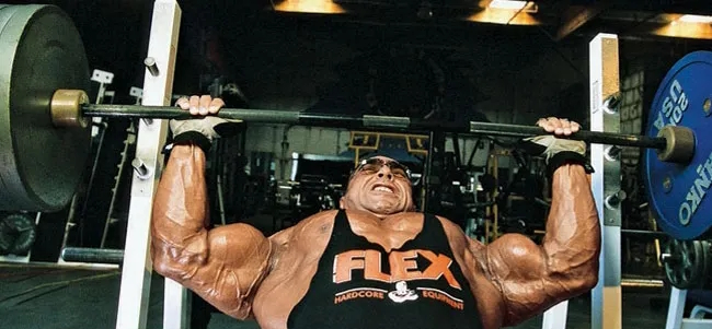
Disclaimer. The material below is intended for a general audience and reflects basic medical principles, but the rules are substantially different in professional bodybuilding. As a sports physician, endocrinologist, and someone who also competes and trains at the limits of physiological capacity — with a 22,4 inch peak arm (author’s photo in the “basement”) — I understand very well that professionals knowingly accept these risks.
With proper endocrinological monitoring, an understanding of pharmacodynamics, and reasonable control of working weights without unjustified “ego lifting,” appropriate pharmacological support can work not against the athlete but in their favor: it increases collagen synthesis, accelerates the regeneration of microinjuries, and protects the ligament-tendon system from premature wear under heavy loads.
There is a dangerous but extremely widespread stereotype that any active participation in sports automatically guarantees a healthy musculoskeletal system.
In reality, moderate physical activity (swimming, therapeutic exercise, elliptical training) does nourish cartilage through the diffusion of synovial fluid, but professional strength sports — powerlifting, weightlifting, and bodybuilding — operate according to completely different biomechanical principles. Cartilage has no intrinsic blood supply and functions like a dense sponge: under moderate compression, it expels used fluid; when the pressure is released, it absorbs fresh nutrient-rich fluid.
When an athlete regularly squats with a 200–300 kg barbell or performs heavy leg presses on a machine, enormous axial compression literally flattens hyaline cartilage into a thin film, blocking its nutrition and causing microtears in the matrix.
The fundamental problem for strength athletes lies in the deep imbalance between the enormous potential of a trained muscular system and the sharply limited reserve strength of connective tissue.
Muscles hypertrophied and stimulated through years of training can generate pulling forces that the passive structures of cartilage, ligaments, and tendons simply are not evolutionarily adapted to withstand. The anatomical capacity of joint surfaces has a genetic limit, and regular work with submaximal weights leads to catastrophically accelerated wear of the cushioning material. Joint tissue becomes thinner many times faster than in a person leading an ordinary lifestyle, turning elite-level sports into a continuous production line for early severe osteoarthritis.
The situation is pushed to a critical point by pharmacological support and the specific competitive nature of elite athletes.
The use of anabolic steroids allows muscles to grow and strengthen at astronomical rates, but these hormones do not strengthen articular cartilage; instead, they increase the fatal gap between muscular pulling strength and the structural strength of the joint. In addition, professional athletes become accustomed to ignoring early pain signals and masking them with heavy doses of nonsteroidal anti-inflammatory drugs. By starting a heavy set under the effects of powerful painkillers, the athlete forcibly shuts down the nervous system’s protective reflexes and systematically destroys the injured joint, grinding its surfaces into powder.
The history of professional bodybuilding is full of examples in which the pursuit of extreme muscular size ended on an orthopedic operating table.
The most illustrative and well-known case is eight-time “Mr. Olympia” Ronnie Coleman. His legendary, unmatched strength workouts, including squats with weights approaching 400 kilograms, led to severe bilateral hip osteoarthritis. Coleman ultimately underwent a series of complex surgeries, including bilateral total hip replacements, as well as multiple surgical procedures on his severely damaged spine. Despite his massive muscular frame, his cartilage simply could not withstand years of extreme compression.
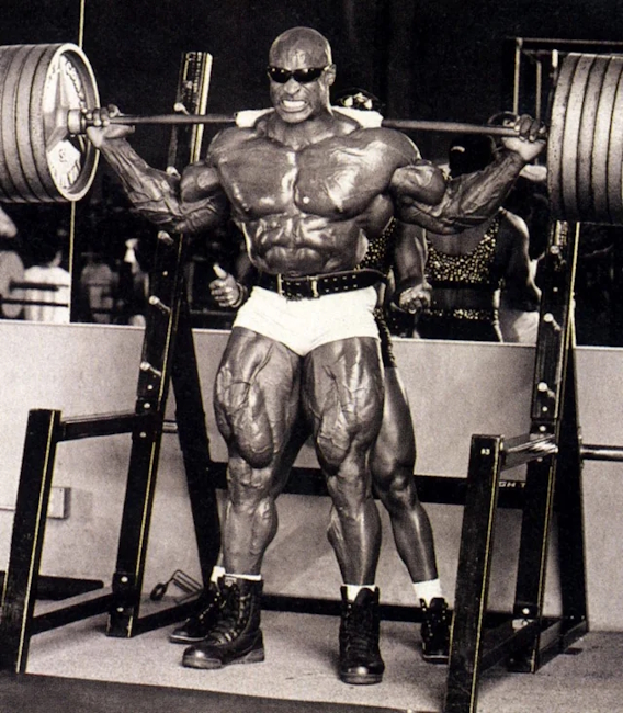
Other elite representatives of the iron sports followed a similar path.
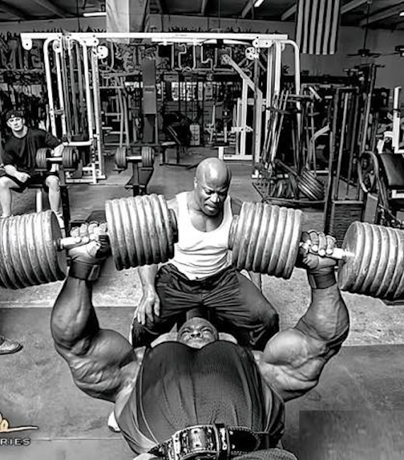
Branch Warren (pictured below), known for his deliberately brutal training style and enormous working weights, repeatedly suffered not only tendon ruptures but also severe wear of his joints, which likewise required major surgical reconstruction.

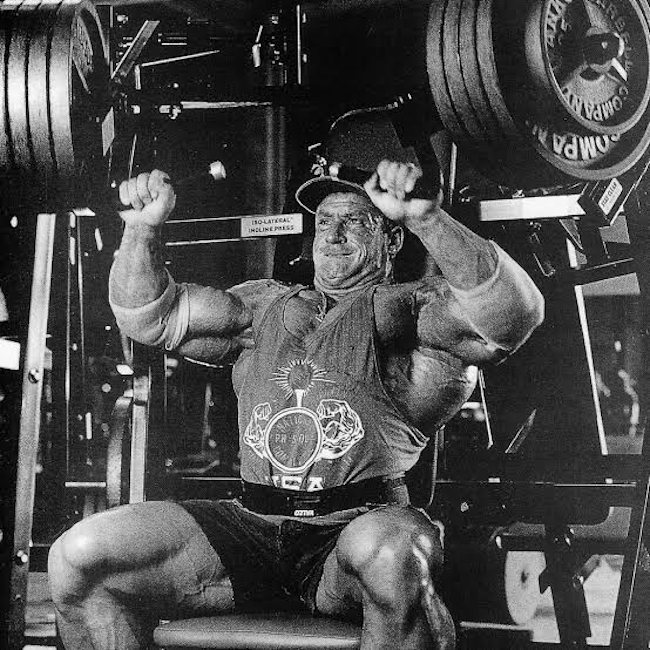
Dorian Yates, a six-time Olympia winner and leading popularizer of high-intensity training to failure with maximal weights, pushed his musculoskeletal system into a state of profound deterioration before his career was over. Their clinical histories convincingly demonstrate that enormous muscle mass does not protect against osteoarthritis when training is consistently built around extreme overloads that exceed the physiological capabilities of the human skeleton.
Now, regarding pharmacology.
Within strength sports, the belief is widespread that anabolic steroids, particularly nandrolone, can restore the joints.
However, this therapeutic effect is largely illusory: nandrolone causes pronounced fluid retention in tissues and the synovial capsule, creating temporary additional cushioning and masking joint pain. Although it can moderately stimulate collagen synthesis, water retention only relieves discomfort without regenerating destroyed hyaline cartilage.
Once the athlete stops feeling pain, they continue training with heavy weights, which means that after the drug is discontinued and excess fluid is lost, the degenerative changes in the joint are discovered in an even more advanced form.
Unlike steroids, growth hormone (somatotropin) and specialized peptide compounds (such as BPC-157 and TB-500) actually stimulate regenerative processes in connective tissue by activating insulin-like growth factor 1 (IGF-1) and accelerating collagen synthesis.
Peptide preparations promote angiogenesis — the formation of new microvessels in ligaments and tendons — which improves nutrition in areas that normally have poor blood supply and accelerates healing of microdamage. Nevertheless, their actual potential is limited to the ligamentous apparatus and early injuries: they cannot fully restore cartilage worn away by Stage 2–3 osteoarthritis, while uncontrolled use of growth hormone additionally carries risks of insulin resistance, sodium retention, and the development of entrapment syndromes.
In summary, it should be emphasized once again (see the Disclaimer at the beginning of the chapter) that the use of nandrolone and growth hormone during high-volume bodybuilding can nourish and protect the joint-ligament system very effectively. The key is not to turn training into powerlifting: avoiding extreme weights allows pharmacology to fully restore tissue instead of driving it toward overuse.

Dmitry Volkov – is the author of our bodybuilding section is a practicing sports medicine physician based in Dallas, Texas, with 21 years of hands‑on experience in sports pharmacology. At 42, he combines deep academic knowledge with real‑world expertise gained from coaching athletes of all levels — from amateurs to seasoned competitors. He earned his medical degree from a leading Texas institution and spent years working in sports medicine clinics and private practice.
His primary focus is hormonal regulation of muscle growth, the use of anabolic steroids and peptides, and post‑cycle recovery. He understands modern protocols inside out because he consults real people every day, helping them avoid side effects and achieve safe results. His approach is rooted in evidence‑based medicine, yet remains grounded in the realities of both amateur and professional sports.
In his articles, he aims to debunk myths and deliver clear, scientifically sound recommendations. Every piece of content is vetted not only by medical knowledge but also by years of clinical observation. He firmly believes that responsible pharmacology requires a solid grasp of biochemistry, respect for one’s body, and regular medical monitoring — and he works hard to convey these principles in a way that is both accessible and actionable for his readers.